Sunday, February 18, 2024

TRANSCRIPTION

 TRANSCRIPTION IN PROKARYOTES

  • Transcription is the process by which the information in a strand of DNA is copied into a new molecule of messenger RNA (mRNA).
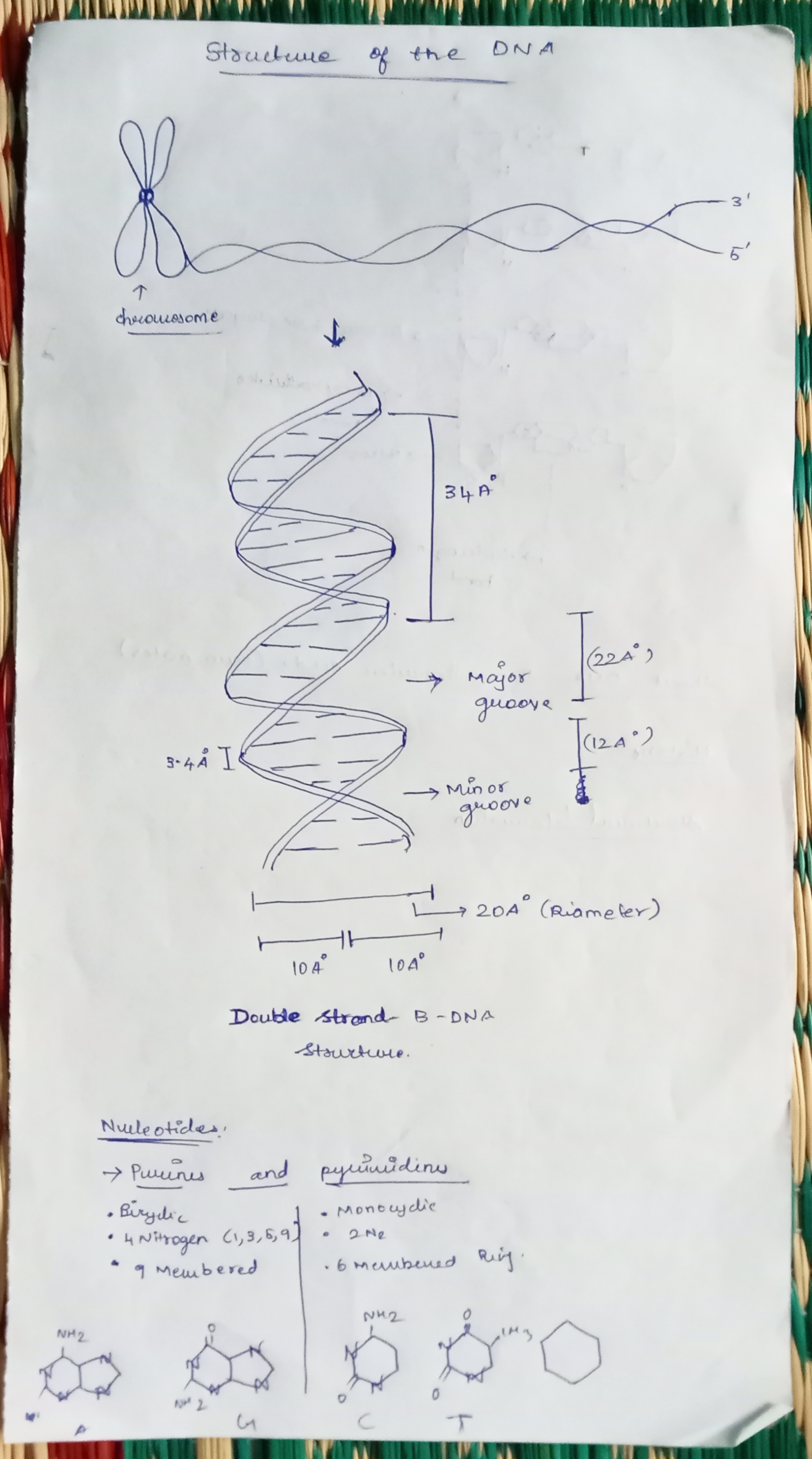
  • During replication entire genome is copied but in transcription only the selected portion of genome is copied.
ENZYME INVOLVED IN TRANSCRIPTION


  • RNA is synthesized by a single RNA polymerase enzyme which contains multiple polypeptide subunits.
  • In E. coli, the RNA polymerase has subunits: two α, one β, one β’ and one ω and σ subunit (α2ββ’ωσ). This complete enzyme is called as the holoenzyme.
  • The σ subunit may dissociate from the other subunits to leave a form known as the core enzyme.
STEPS INVOLVED IN PROKARYOTIC TRANSCRIPTION

Transcription is an enzymatic process. the mechanism of transcription completes in three major steps

1. Initiation:

  • closed complex formation
  • Open complex formation
  • Tertiary complex formation

2. Elongation

3. Termination:

INITIATION:

  • The transcription is initiated by RNA polymerase holoenzyme from a specific point called promoter sequence.
  • Bacterial RNA polymerase is the principle enzyme involved in transcription.
  • The core enzyme bind to specific sequence on template DNA strand called promotor. The binding of core polymerase to promotor is facilitates and specified by sigma (σ) factor. (σ70 in case of E. coli).
  • The core polymerase along with σ-factor is called Holo-enzyme ie. RNA polymerase  holoenzyme.
  • In case of e. coli, the promoter consists of two short sequences at -10 and -35 positions upstream from the transcription start site.The sequence at -10 is called the Pribnow box, or the -10 element, and usually consists of the six nucleotides TATAAT. The Pribnow box is absolutely essential to start transcription in prokaryotes.The other sequence at -35 (the -35 element) usually consists of the six nucleotides TTGACA. Its presence allows a very high transcription rate.

i. closed complex:

  • Binding of RNA polymerase holoenzyme to the promotor sequence form closed complex
ii. Open Complex: 

  • After formation of closed complex, the RNA polymerase holoenzyme separates 10-14 bases extending from -11 to +3 called melting. So that open complex is formed. This changing from closed complex to open complex is called isomerization.
iii. Closed Complex: 


  • RNA polymerase starts synthesizing nucleotide. It does not require the help of primase.
  • If the enzyme synthesize short RNA molecules of less than 10 bp, it does not further elongates which is called abortive initiation. This is because σ 3.2 acting as mimic of RNA and it lies at middle of RNA exit channel in open complex.
  •   When the RNA polymerase manage to synthesize RNA more than 10 bp long, it eject the σ 3.2 region and RNA further elongates and exit from RNA exit channel. This is the formation of tertiary complex.

ELONGATION

The transcription elongation phase begins with the release of the σ subunit from the polymerase. The dissociation of σ allows the core RNA polymerase enzyme to proceed along the DNA template, synthesizing mRNA in the 5′ to 3′ direction at a rate of approximately 40 nucleotides per second. As elongation proceeds, the DNA is continuously unwound ahead of the core enzyme and rewound behind it. Since the base pairing between DNA and RNA is not stable enough to maintain the stability of the mRNA synthesis components, RNA polymerase acts as a stable linker between the DNA template and the nascent RNA strands to ensure that elongation is not interrupted prematurely. The synthesized RNA is proof reads by Hydrolytic editing. For this the polymerase back track by one or more nucleotide and cleave the RNA removing the error and synthesize the correct one. The Gre factor enhance this proof reading process.


TERMINATION

There are two mechanism of termination.

i) Rho - independent:

  • In this mechanism, transcription is terminated due to specific sequence in terminator DNA.
  • The terminator DNA contains invert repeat which cause complimentary pairing as transcript RNA form hair pin structure.
  • This invert repeat is followed by larger number of TTTTTTTT(~8 bp) on template DNA. The uracil appear in RNA. The load of hair pin structure is not tolerated by A=U base pair so the RNA get separated from RNA-DNA heteroduplex.


Rho - Dependent termination: 

  • In this mechanism, transcription is terminated by rho (ρ) protein.
  • It is ring shaped single strand binding ATpase protein.
  • The rho protein bind the single stranded RNA as it exit from polymerase enzyme complex  and hydrolyse the RNA from enzyme complex.
  • The rho protein does not bind to those RNA whose protein is being translated. Rather it bind to RNA after translation.
  • In bacteria transcription and translation occur simultaneously so the rho protein bind the RNA after translation has completed but transcription is still ON.       


 TRANSCRIPTION IN EUKARYOTES

  • Transcription is the process by which the information in a strand of DNA is copied into a new molecule of RNA.
  • It is the first step of gene expression, in which a particular segment of DNA is copied into RNA (especially 
  • 'RNA) by the enzyme RNA polymerase.

ENZYME INVOLVED IN THE EUKARYOTIC TRANSCRIPTION

  • RNA polymerase I (RNA Pol I) is located in the nucleolus and transcribes the 28S, 18S, and 5.8S rRNA genes.
  • RNA polymerase II (RNA Pol II) is located in the nucleoplasm and transcribes protein-coding genes, to yield pre-mRNA, and also the genes encoding small nucleolar RNAs (snoRNAs) involved in rRNA processing and small nuclear RNAs (snRNAs) involved in mRNA processing, except for U6 snRNA.
  • RNA polymerase III (RNA Pol III) is also located in the nucleoplasm. It transcribes the genes for tRNA, 5S rRNA, U6 snRNA, and the 7S RNA associated with the signal recognition particle (SRP) involved in the translocation of proteins across the endoplasmic reticulum membrane.
  • Each of the three eukaryotic RNA polymerases contains 12 or more subunits and so these are large complex enzymes.
  • The genes encoding some of the subunits of each eukaryotic enzyme show DNA sequence similarities to genes encoding subunits of the core enzyme of E. coli RNA polymerase.
  • However, four to seven other subunits of each eukaryotic RNA polymerase are unique in that they show no similarity either with bacterial RNA polymerase subunits or with the subunits of other eukaryotic RNA polymerases.

PROCESS OF EUKARYOTIC TRANSCRIPTION

The basic mechanism of RNA synthesis by these eukaryotic RNA polymerases can be divided into the following phases:

a) INITIATION PHASE: 

  • PAPER 
b) ELONGATION PHASE:

TFIIH has two functions:

  1. It is a helicase, which means that it can use ATP to unwind the DNA helix, allowing transcription to begin.
  2. In addition, it phosphorylates RNA polymerase II which causes this enzyme to change its conformation and dissociate from other proteins in the initiation complex.
  • The key phosphorylation occurs on a long C-terminal tail called the C-terminal domain (CTD) of the RNA polymerase II molecule.
  • Interestingly, only RNA polymerase II that has a non-phosphorylated CTD can initiate transcription but only an RNA polymerase II with a phosphorylated CTD can elongate RNA.
  • RNA polymerase II now starts moving along the DNA template, synthesizing RNA, that is, the process enters the elongation phase.
  • TF II S is used to increase the speed of the transcription process. 
  • RNA synthesis occurs in the 5’ → 3’ direction with the RNA polymerase catalyzing a nucleophilic attack by the 3-OH of the growing RNA chain on the alpha-phosphorus atom on an incoming ribonucleoside 5-triphosphate.
  • The RNA molecule made from a protein-coding gene by RNA polymerase II is called a primary transcript.
TERMINATION:

(PAPER)


Tuesday, February 13, 2024

RESTRICTION ENZYMES

RESTRICTION ENZYMES

A restriction enzyme is a nuclease enzyme that cleaves DNA sequence at a random or specific recognition sites known as restriction sites. In bacteria, restriction enzymes form a combined system (restriction + modification system) with modification enzymes that methylate the bacterial DNA. Methylation of bacterial DNA at the recognition sequence typically protects the own DNA of the bacteria from being cleaved by restriction enzyme

There are two different kinds of restriction enzymes:

 (1) Exonucleases catalyses hydrolysis of terminal nucleotides from the end of DNA or RNA molecule either 5’to 3’ direction or 3’ to 5’ direction. Example: exonuclease I, exonuclease II etc. 

(2) Endonucleases can recognize specific base sequence (restriction site) within DNA or RNA molecule and cleave internal phosphodiester bonds within a DNA molecule. Example: EcoRI, Hind III, BamHI etc

EXONUCLEASES AND IT TYPES: 


EXONUCLEASE III: 

  • Exonuclease III is a globular enzyme which has 3’→5’ exonuclease activity in a double stranded DNA. 
  • The template DNA should be double stranded and the enzyme does not cleave single stranded DNA. 
  • The enzyme shows optimal activity with blunt ended sequences or sequences with 5’ overhang. 
  • Exonuclease III enzyme has a bound divalent cation which is essential for enzyme activity.
  • The mechanism of the enzyme can be affected by variation in temperature, monovalent ion concentration in the reaction buffer, and structure and concentration of 3’termini. 
  • The enzyme shows optimal activity at 37°C at pH 8.0.

MUNG BEAN NUCLEASES:

  • As the name suggest, this nuclease enzyme is isolated from mung bean sprouts (Vigna radiata). 
  • Mung bean nuclease enzymes can degrade single stranded DNA as well RNA. Under high enzyme concentration, they can degrade double stranded DNA, RNA or even DNA/RNA hybrids.
  •  Mung bean nuclease can cleave single stranded DNA or RNA to produce 5’-phosphoryl mono and oligonucleotides.
  • It requires Zn2+ ion for its activity and shows optimal activity at 37°C. 
  • The enzyme works in low salt concentration (25mM ammonium acetate) and acidic pH (pH 5.0). 
  • Treatment with EDTA or SDS results in irreversible inactivation of the enzyme. Mung bean nuclease is less robust than S1 nuclease and easier to handle. It has been used to create blunt end DNA by cleaving protruding ends from 5’ ends. This enzyme cannot produce nicks in a double stranded DNA but at higher concentration, it can generate nicks and cleave double stranded DNA.


RNases: 

Ribonucleases are a class of hydrolytic enzymes that catalyzes both the in vivo and in vitro degradation of ribonucleic acid (RNA) molecules into smaller components. Ribonucleases are classified into two types, Exoribonucleases and endoribonucleases. 

Exoribonucleases: The exoribonuclease is an exonuclease ribonuclease that degrades RNA by removing terminal nucleotides from either the 5′ end or the 3′ end of the RNA molecule.

Endoribonucleases: The endonuclease ribonuclease cleaves RNA molecules internally. It can cleave either single-stranded or double-stranded RNA, depending on the enzyme.

RNases I:

RNases A:

RNases H:


T4 PHOSPHO NUCLEOTIDE KINASE:

Polynucleotide kinases (PNK) are one such DNA modification enzymes that add phosphate groups to nucleic acid molecules. They transfer the gamma phosphate group from adenosine triphosphate (ATP) to the 5’ hydroxyl termini of DNA or RNA.

                        ATP + 5'-dephospho-DNA  ADP + 5'-phospho-DNA

Thus, the two substrates of this enzyme are ATP and 5'-dephospho-DNA, whereas its two products are ADP and 5'-phospho-DNA. Polynucleotide kinase is a T7 bacteriophage (or T4 bacteriophageenzyme that catalyzes the transfer of a gamma-phosphate from ATP to the free hydroxyl end of the 5' DNA or RNA. The resulting product could be used to end-label DNA or RNA, or in ligation reactions.

DUAL ROLE OF T4 Polynucleotide Kinase

The dual roles—kinase and phosphatase—performed by the enzyme reside in its separate domains. The N-terminal is a 5′-kinase domain with a nucleotide-binding motif GXXXXGK(S/T) and a C-terminal is a 3′-phosphohydrolase domain.


TYPES OF RESTRICTION ENZYMES

Restriction enzymes are traditionally classified into four types on the basis of subunit composition, cleavage position, sequence specificity and cofactor requirements. However, amino acid sequencing has uncovered extraordinary variety among restriction enzymes and revealed that at the molecular level, there are many more than four different types.

TYPE 1

Type I restriction enzymes (REases) are large pentameric proteins with separate restriction (R), methylation (M) and DNA sequence-recognition (S) subunits. They were the first REases to be discovered and purified, but this type is not used as like as Type 2. 

Enzyme Activity:

Type I R–M enzymes are pentameric proteins of composition 2R+2M+S. They require ATP, Mg2+ and S-adenosylmethionine (SAM) for activity and display both REase and MTase activities. A trimer of 2M+S acts solely as an MTase 

Friday, January 26, 2024

mRNA in Prokaryotic and Eukaryotic cell

RIBONUCLEIC ACID - mRNA (Prokaryotic and Eukaryotic)

  • mRNA accounts for 5% of the total RNA. A ribose nucleotide in the chain of RNA consists of a ribose sugar, phosphate group, and a base. 
  • In each ribose sugar, one of the four bases is added: Adenine (A), Guanine (G), Cytosine (C), and Uracil (U).
  • The base is attached to a ribose sugar with the help of a phosphodiester bond. As RNA comprises many ribose nucleotides, the length of the chains of nucleotides can vary according to their types or their functions.
  • RNA thus differs from DNA, on the type of sugar used to make the molecule and replacement of base Thymine in DNA with Uracil in RNA. Additionally, DNA is a double-stranded molecule whereas RNA is a single-stranded molecule.
  • ·   It carries genetic information in the form of triplet codon.
  • START CODON - AUG  
  • STOP CODON   - UAA, UGA, UAG.


PROKARYOTIC mRNA


·   mRNA is a single-stranded RNA molecule running from 5’ to 3’ direction.

·   It has sort lifespan and less stable.

·   In a prokaryotic cell, there is a lack of a distinct nucleus, thus the mRNA synthesized contained a copy of DNA sequences with a terminal 5’- triphosphate group and 3’ hydroxyl group. Prokaryotic mRNA at its 5’ end has a Shine Dalgarno sequence which is rich in purine nucleotide and is essential for facilitating the binding of mRNA to ribosome.

·   Prokaryotic mRNA starts at 5’ with a triphosphate group, followed by Shine Dalgarno sequences (untranslated region) and as we move along to the 3’ direction, the coding regions begin. The coding regions begin with a start codon and end when it reaches a stop codon, after encountering a stop codon, protein-coding regions come to an end, after which there is another untranslated region that terminates at the end of 3’.

·   This prokaryotic mRNA is polycistronic in nature.

·   It has Operon (Cluster of genes).

·   Translation and transcription take place in cytoplasm.

·   mRNA processing is not seen in prokaryotes.


EUKARYOTIC mRNA

  • However, in a eukaryotic cell, the nucleus is distinct and has numerous enzymes in it that make the synthesized mRNA molecule go through modification at their terminals 5’ and 3’ to maintain the integrity and stability of the mRNA molecule (post-transcriptional modification).
  • The 5’ triphosphate group of eukaryotic mRNAs is esterified forming a cap structure. This process is referred to as 5’ capping, which happens when a 7-methylguanosine cap is added to a 5’ free triphosphate group via 5’-5’ phosphate linkage with the help of an enzyme called guanyl transferase.
  • The 5’ capping is important for the recognition of mRNA by the Ribosomes during protein synthesis. 
  • And likewise, the 3’ hydroxyl group is cleaved to give a free hydroxyl group to which numerous adenine monophosphates are added to make the mRNA molecule more stable and prevent it from degradation. This tail of numerous adenine nucleotides is referred to as Poly-A-tail.
  • The Poly-A-tail added to the 3’ end of eukaryotic mRNA are usually 100-200 bases long; the addition is catalyzed by an enzyme poly(A) polymerase that recognizes the sequence AAUAAA as a single for addition.
  • The poly-A-tail is vital during transporting mRNA from the nucleus to the cytoplasm for proteins synthesis.
  • Eukaryotic mRNA starts at 5’ with a cap followed by untranslated regions (5’ UTR), and as we move along 3’ direction, the coding regions begin with the start codon and continue till it reaches the stop codon, which marks the termination of coding regions, after which there is another untranslated region (3’ UTR) that terminate when Poly-A-tail begins, which further terminates at the end of 3’.

  • UTR – Untranslated region sis mRNA domain, it contains regulatory elements which have two types, Cis regulatory elements, Trans – regulatory elements. The CRE elements present I the nearby location of the genes that are going to regulate the genes by acting as a Promotor (initiating transcript), Enhancer (Enhance the transcriptase), Silencer (Inhibit the Transcript).   &   The Trans Elements located at different location of the genome to the gene that they regulate. They are regulating by the repressor molecule.

  • The 3′-untranslated region plays a crucial role in gene expression by influencing the localization, stability, export, and translation efficiency of an mRNA. It contains various sequences that are involved in gene expression, including microRNA response elements (MREs), AU-rich elements (AREs), and the poly(A) tail. In addition, the structural characteristics of the 3′-UTR as well as its use of alternative polyadenylation play a role in gene expression.


Sunday, January 14, 2024

ABOUT DNA AND ITS STRUCTURE

                                              STRUCTURE OF THE DNA 


DEFINITION 

     DNA, or deoxyribonucleic acid, is the hereditary material in humans and almost all other organisms. Nearly every cell in a person’s body has the same DNA. Most DNA is located in the cell nucleus (where it is called nuclear DNA), but a small amount of DNA can also be found in the mitochondria and Chloroplast. The information in DNA is stored as a code made up of four chemical bases: adenine (A), guanine (G), cytosine (C), and thymine (T). Human DNA consists of about 3 billion bases, and more than 99 percent of those bases are the same in all people.  The genome of the organism consists of Introns(Non coding genes)  and Exons (coding region). And only 2% of the total genome codes for protein, and others are intron (which is non - coding genes). 


HISTORY

     In the 1950s, Francis Crick and James Watson worked together at the University of Cambridge, England, to determine the structure of DNA.   In Wilkins’ lab, researcher Rosalind Franklin was using X-ray crystallography to understand the structure of DNA. Watson and Crick were able to piece together the puzzle of the DNA molecule using Franklin’s data.  Watson and Crick also had key pieces of information available from other researchers such as Chargaff’s rules. Chargaff had shown that of the four kinds of monomers (nucleotides) present in a DNA molecule, two types were always present in equal amounts and the remaining two types were also always present in equal amounts. This meant they were always paired in some way.


STRUCTURE OF THE DNA


a)   The building blocks of DNA are nucleotides, which are made up of three parts: a deoxyribose (5-carbon sugar), a phosphate group, and a nitrogenous base. 
* Deoxyribose Sugar Structure 
* N2 Bases Structure 
* Bonds: Hydrogen Bond, Phosphodiester bond

b) KEY POINTS: 
DNA is made of two helical chains that intertwine with each other to form a double helix. The most widely accepted structure of DNA is right-handed helix DNA also known as the B-form of DNA, which is 1.9 nm in diameter.
These helical chains run anti-parallel to each other, one polynucleotide chain runs from 5’ to 3’ and the other polynucleotide chain runs from 3’ to 5’. These chains are connected to each other via nitrogen bases through hydrogen bonding.
Hydrogen bonding contributes to the specificity of base pairing. Adenine preferentially pairs with Thymine through 2 hydrogen bonds. Similarly, Cytosine preferentially pairs with Guanine through 3 hydrogen bonds. 
We can even say, that the base pairing happens when Pyrimidines pair with Purines because Pyrimidines refers to the single ring structure of Thymine and Cytosine and Purines refers to double-ring structures, Adenine and Thymine.
The base pairs A = T and G ≡ C are known as complementary base pairs. Hence, the amount of Adenine is equal to the amount of Thymine, and the amount of Guanine is equal to the amount of Cytosine.
The geometry of the DNA is influenced by the distance between the backbones and the angle at which the nitrogenous bases are attached to the backbone.
The major groove occurs when the backbones are far apart from each other and the minor groove occurs when they are close.
The regularity of the helical structure forms two repeating and alternating spaces: Major and Minor grooves.
These groves act on base-pair recognition and binding sites for protein, the major groove contains base pair specific information while the minor groove is largely base-pair nonspecific, caused by protein interactions in the grooves
The double-helical structure of DNA is highly regular, each turn of the helix measures approximately 10 base pairs. In addition to hydrogen bonding in between the bases, the staging of bases also stabilizes the structure, there are pi-pi interactions between staged aromatic rings of the bases.
The distance between each turn is 3.4 nm.
The major groove is 2.2 nm wide and the minor groove is 1.1 nm wide.
Chargaff's Rule: Chargaff's rules state that DNA from any cell of all organisms should have a 1:1 ratio (base Pair Rule) of pyrimidine and purine bases and, more specifically, that the amount of guanine is equal to cytosine and the amount of adenine is equal to thymine.


Monday, December 25, 2023

ANTI - CANCER EFFECTS OF PLANT SECONDARY METABOLITES AGAINST VARIOUS CANCER


1. INTRODUCTION

     Secondary metabolites are not essential for the growth and development of plants, but they have important accessory activities such as help in defence against herbivory, growth inhibition of competing plants, bacterial and fungal pathogens and aiding in pollination. Notable anticancer alkaloids include vinblastine, vincristine and camptothecin; terpenoids include lycopene and gamma-tocopherol; polyphenols include etoposide, resveratrol, curcumin and epigallocatechin gallate (EGCG); and flavonoids include apigenin, genistein and kaempferol. These bioactive compounds exert anticancer effects either independently or synergistically with other compounds through regulation of metabolic and signalling pathways, inhibition of enzymes vital for cancer progression, angiogenesis, microtubule assembly and inducing apoptosis.  


2. ANTI - CANCER EFFECTS OF PLANT SECONDARY METABOLITES

     The hallmark of a cancer cell is its uncontrolled rate of proliferation. A cancer cell alters its physiology to meet the nutritional and energy requirements. In case of scarcity of nutrients and energy, cancer cells are able to modulate their pathways and continue proliferation. These alteration are in the form of modification in metabolic pathways, signalling pathways an enzymatic regulation.  These metabolic alteration mainly focus on rapid ATP production, synthesis of macromolecules needed for cell progression and regulation of appropriate redox state. 

     By understanding those pathways, researchers used different secondary metabolites from plants against Various Lung Cancer.

2.1. ANTICANCER EFFECT OF NARINGENIN AGAINST BREAST CANCER

In 2015, Using the concept of Warn burg Effect, throne and Campbell it has been reported that glucose transporters (GLUT) are up - regulated in cancer in a cell - specific manner. (The Warburg and colleagues made the observation that tumour cells produce energy predominantly not through the usual citric acid cycle and oxidation phosphorylation in the mitochondria as observed in normal cells, but through a less efficient process of 'aerobic glycolysis' consisting of high level of glucose uptake and glycolysis followed by lactic acid fermentation).  Target GLUT presents a viable strategy for cancer inhibition and treatment. Plant extract have been shown to target GLUT. For example, naringenin, a flavonoid presents in grapes inhibits glucose uptake in MCF- 7 Breast cancer cell by inhibiting the phosphoinositide 3 - kinase (PI3K) pathway that regulate glucose transporter, GLUT 4. 


2.2. ANTICANCER EFFECTS OF DELPHINDIN AGAINST NON SMALL CELL LUNG CANCER

In 2017, Kim Ed et al, reported the antiangiogenic activity of delphinidin. Delphinidin is a flavonoid which is abundantly found in fruits, flowers and leaves of plants.  They found that delphinidin decreases the expression level of HIF - 1, which is a VEGF transcription factor. (VEGF - Vascular Endothelial Growth Factor is a homodimer glycoprotein, it is main key factor in the process of making new blood Vessels. These Vascular endothelium, a monolayer of endothelial cells, Constitutes the inner cellular lining of arteries, Veins and Capillaries and therefore is in direct contact with the components and cells of blood).  


ADVANTAGES 

 The Production of secondary metabolites from plant cells via in vitro techniques under aseptic conditions as the potential of providing an unlimited supply of targeted compounds. The PTC is not used to synthesize the Anticancer compounds but also biopharmaceuticals including therapeutics antibodies for other diseases. The advantage of the callus culture, is we can maintain the cell at the stationary phase by providing the unlimited supply of the target secondary compounds. 

  • Because of their targeted action, these drugs have an effect on the cancer cells and mostly leave normal, healthy cell alone. Traditional chemotherapy is cytotoxic to most cells, meaning it can damage normal, healthy cells in addition to damaging and killing cancer cells. 
  • For the Cancer stages, I, IIA, IIB, IIIB, IIIC, the target therapy is not need, but the Stage IV treatment which includes Target therapy with Angiogenesis inhibitor, Target therapy with Apoptosis Initiator. 

DISADVANTAGES

                                                 RESEARCH CARRIED OUT BY IIT MADRAS
  • They collect the plant which synthesize the cyclopeptides as a secondary metabolites, which is used as target molecule against the cancer cells. 
  • They collect from the Himalayas and the ooty region, then they cultivated that in the horticulture in IIT madras. After 3 years they begin the study by characterizing the different types of cyclopeptides in the plants.

  • RESULT:  What they found is the plant species from Himalaya and ooty, which was cultivated in the IIT Madras, they show Different array of Peptides, and its also synthesize the new proteins which are not in the plants in the Himalayas. This study which concludes, there is high impact of Climatic and Geographical Variabilities. 

OVERVIEW OF NON - SMALL CELL LUNG CANCER


INTRODUCTION

Lung Cancer is the leading cause of Cancer related death worldwide, and approximately 85% of cases are related to cigarette smoking. Metastasis, which is common in lung cancer is a multi stage process, involved invasion into blood or lymph, extravasation and growth at a new site. This Blog gives a way to understand what is Non Small Cell Lung Cancer, and its types and stages and How they are treated using the Secondary Metabolites from the living organisms (Particularly and Plants).


ABOUT NSCLC

  • NSCLC is the most common type  of lung Cancer. It comprises 80 - 85% of lung Cancer. 
  • This NSCLC is a malignant tumour with extremely high Mortality.
  • NSCLC, it name implies that the size of the cancer cell is larger than the Small cell Lung cancer cell, by viewed under the microscope.
  • NSCLC is the common type of Lung Cancer.
  • It stages from 0 - IV. It mean, we can classify the stage of the NSCLC, based on the 
  • Because of the larger  in size, it grow and spread slowly compared to the SCLC.
  • The most common subtype of NSCLC are: 
a) Lung Adenocarcinoma: This is a slow growing subtype. It often develops in the bronchi or airways of the lungs. 
b) Lung Squamous cell Carcinoma: This is the least common type of NSCLC. It can form in any part of the lung and tends to grow and spread rapidly.  
c) Large cell carcinoma: This  is the most common type of NSCLC. It typically begins in the outer tissues of the lung that secrete substances such as mucus. This type  is more likely to be diagnosed before it spreads. 

HOW THE NSCLC IS CAUSED

Tobacco smoking is the leading cause of cancer - related death in the world, having been associated to approximately 1.2 million death annually, and it is linked 90% of lung cancer cases. We can classify the Causes into 3 forms, Based on How we exposed to them: 

A) USING TOBACCO:

There are approximately more than 500 to 600 substances used in cigarette. When we burned cigarettes, it create more than 6000 chemicals. From those chemicals, 60 to 70 chemicals are known to cause cancer. Those chemical cancer causing substances are derived from various chemical classes such as Polycyclic aromatic hydrocarbons (PAHs), Nitrosamines, Aromatic Amines, phenols, volatile hydrocarbons, etc., Example: Acetone, Nicotine, Tar, Toluene are the example of the chemicals in tobacco smoke. 

B) SECONHAND TOBACCO SMOKE

Non - Smokers can be exposed to second-hand smoke from different sources, such as in the home, the workplace, and outside public building. Side stream smoke and mainstream second - hand smoke are different in their physiochemical Properties. The ration of side stream to mainstream smoke vary largely depending on the constituents of tobacco products from different manufactures. For example, nicotine, NNK, and NNN ratio can be 7.1, 0.40, and 0.43 respectively.    

C) SMOKE LESS TOBACCO


Smokeless tobacco is unburned tobacco, and is also known as chewing tobacco, oral tobacco, split or splitting tobacco, dip and snuff. Users chew or suck the tobacco in their mouth and split out the juice of the tobacco. However, a study by Hecht et.al., (2017) demonstrates that there is similar exposure to the tobacco - specific carcinogen NNK in smokers and smokeless tobacco users. 


MECHANISM OF NSCLC DEVELOPMENT:

EXAMPLE:  HOW NITROSAMINE COMPOUND INDUCING CANCER

Naturally occurring in tobacco smoke is a procarcinogen, an inert form that require metabolic activation to exert its carcinogenic functions. The Cytochrome P450 usually metabolizes carcinogens by converting the chemicals to more potent carcinogens, by activating the NNK (Nitrosamine compounds) into DNA reactive metabolites that can induce the methylation, pyridyloxobutylation and pyridylhdroxybutylation of nucleobases in DNA and form DNA adducts. The Repair mechanism either should repair the DNA adducts or apoptosis, if its fails it leads to Mutation in multiple genes and finally cancer. 

STAGES OF NON SMALL CELL LUNG CANCER

STAGE 0 - Non Invasive pre - cancer found in lining of airways.
 
STAGE 1 -  Invasive tumour hasn't spread to lymph nodes. 

STAGE 2 - Tumour is 3 to 7 cm and has spread to nearby tissues or lymph nodes, but not to distant body parts. 

STAGE 3 - Tumour is 3 to 7 cm and has spread to nearby tissues or lymph nodes, but not to distant body parts. 

STAGE 4 - Cancer has potentially spread outside the lung. 


TREATMENT FOR NON SMALL CELL LUNG CANCER: 

There are two method of Treatments are available for NSCLC. 

LOCAL TREATMENT:


Local treatment which are used to treat a specific tumour or area of the body. Example of the local treatment, like surgery and radiation therapy. 

TARGET THERAPY: 


Target therapy is a type of cancer treatment that uses drug designed to "target" cancer cells without affecting normal cells. Cancer cells typically have changes in their genes that make them different from normal cells. Genes are part of a cell's DNA that tell the cell to do certain things. When a cell has certain gene changes it doesn't behave like a normal cell.  


CONCLUSION: 

This is all about the Non small cell lung Cancer, I hope this will helps you to understand the basic concept of NSCLC, classification, Causes (especially tobacco), Stages and its therapies.